Core expertise
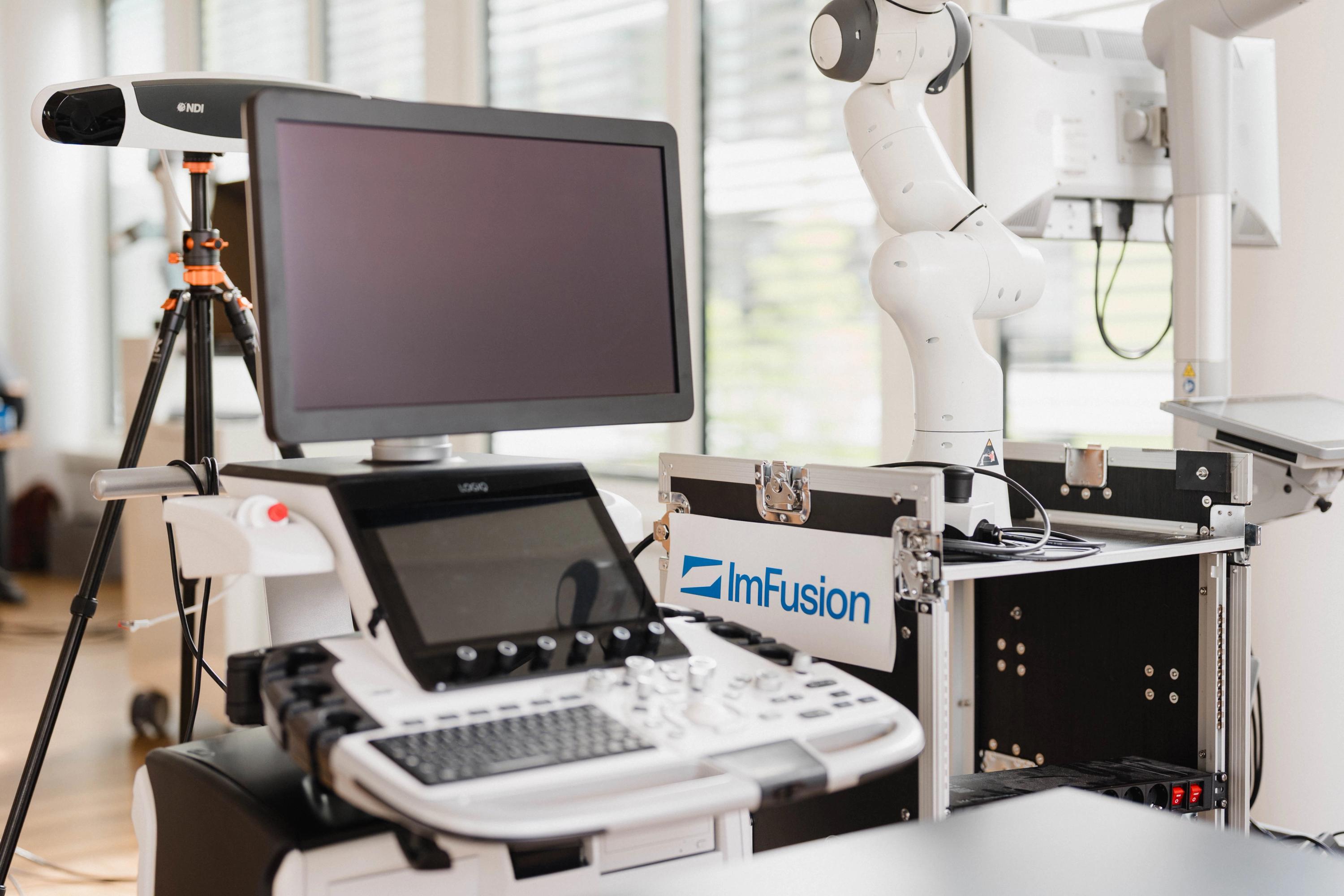
All the expertise your imaging solution needs
Advanced medical imaging requires expertise in algorithms, imaging modalities, real-time processing, and clinical workflows. At ImFusion, we bring these capabilities together in one team to solve challenges across the complete imaging workflow.
Where scientific excellence meets industry experience
Our expertise is backed by a strong track record of scientific publications and successful collaborations with more than 20 industry partners. We apply the latest research to real-world product challenges, turning advanced imaging technologies into practical solutions.
Endoscopic vision
Reliable 3D understanding from endoscopic videos
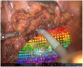
Endoscopic imaging provides live camera views during minimally invasive procedures. We understand its unique challenges, from lens distortion, reflections, and limited depth information to moving cameras, deforming tissue, and instrument occlusion. We combine this domain knowledge with computer vision to reconstruct 3D scenes, track anatomy, and align live video with preoperative data.
3D scene understanding
We understand how to derive depth, camera motion, and anatomical geometry from monocular and stereo endoscopic video. This allows you to transform endoscopic images into spatial information for measurement, mapping, and guidance.
Tissue tracking and anatomical alignment
We combine feature tracking, depth information, and registration methods to follow moving tissue and align preoperative models with live video. This helps you connect surgical plans and segmented anatomy with the intraoperative view.
Phase classification and anatomy segmentation
We have proven models for training surgical phase classification algorithms and anatomy segmentation models based on our customers specific requirements.
Real-time processing and integration
We know how to optimize vision pipelines for live video and integrate them with cameras, computing platforms, and surgical systems, including support for Nvidia Jetson/IGX and Holoscan-based platforms. This enables you to move from individual algorithms to a responsive endoscopic application.
Ultrasound processing
World-renowned expertise in understanding 2D, 3D, and 4D ultrasound images in real-time
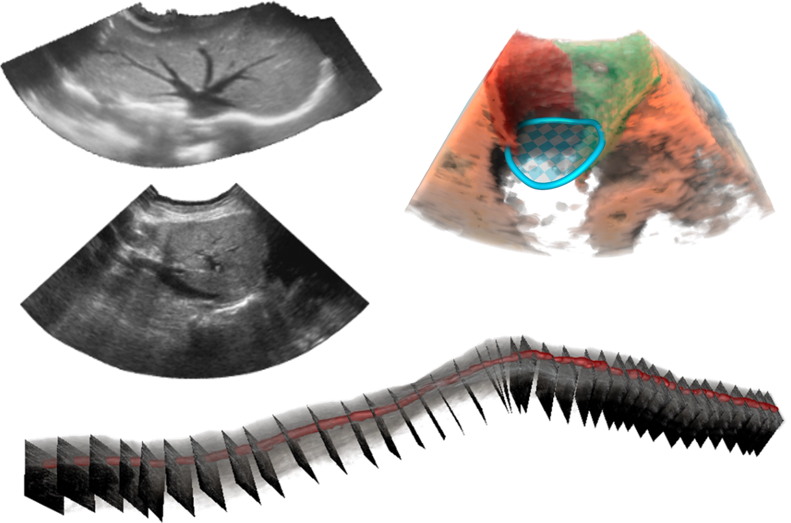
Processing ultrasound images is a core strength of ImFusion. We bridge form any input stage (channel capture to B-mode), extract, segment, reconstruct, and register, and thus allow seamless navigational workflows for any use case.
Seamless hardware integration
We developed algorithms that can robustly deal with differences in image contrast, resolution, field of view, and acquisition geometry. This allows us to align data any modality, including CT, MRI, Ultrasound, X-ray, PET, and endoscopic imaging.
Real-time image analysis
We develop dedicated image filtering, segmentation, detection, and feature extraction workflows with state-of-the-art AI models. Often, these models exploit aspects of ultrasound physics and the inherent modality-specific artifacts.
3D free-hand ultrasound reconstruction
We have been reconstructing 3D ultrasound since 2014 both with and without dedicated tracking equipment. We won the TUS-REC 2025 challenge on trackerless 3D ultrasound and have adopted this technology multiple times for medical devices. Our powerful live compounding algorithms allow on-the-fly reconstruction.
Multi-modal applications
We shape the state-of-art in multi-modal ultrasound registration. Dedicated conventional and AI-based similarity metrics allow for blazingly fast global registrations, even when custom deformation models are involved. We can stitch and decompress, compensate for motion, and even recover probe trajectories from pair-wise acquisitions.
X-ray & CT reconstruction
From projection to precision: GPU-accelerated cone-beam reconstruction and 2D/3D registration for the next generation of X-ray-guided systems
ImFusion brings rich, production-grade expertise in X-ray and CT imaging to teams building the next generation of interventional and diagnostic systems. Our GPU-accelerated software stack takes raw X-ray projections all the way to clinically usable 3D volumes, and precisely fuses live fluoroscopic or C-arm images with pre-operative CT scans or meshes. Built on the same production-grade C++/Python SDK that powers our commercial and research partners' certified products, our X-ray/CT technology is engineered for the accuracy, robustness, and regulatory rigor that medical devices demand.
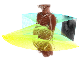
Complete cone-beam CT reconstruction pipeline
We cover the complete reconstruction chain, from projection preprocessing (flat-field correction, log conversion, dead-pixel inpainting) to GPU-accelerated analytical and iterative reconstruction (MLEM, SART, SQS, CG, TV-regularized). Our experience with curved detectors, half-scan, biplanar, and C-arm geometries allows us to adapt the pipeline to your imaging system and application.
Advanced 2D/3D registration and fusion
We combine GPU-based forward projection with multi-resolution intensity registration to align X-ray or fluoroscopy images with CT volumes and anatomical meshes. Different initialization methods and dedicated support for EOS biplanar systems help us address a wide range of clinical setups.
Geometric calibration and artifact reduction
We understand how detector geometry, system configuration, and acquisition conditions affect image quality and can flexibly calibrate for diverse detector types and vendors. Our methods cover C-arm distortion correction, geometric self-calibration, and the reduction of metal, beam-hardening, and ring artifacts.
Physics-based simulation and deep learning readiness
We use GPU-accelerated forward projection, spectral X-ray modelling, and realistic noise simulation to generate representative synthetic images. This provides suitable data for applications such as model training, fiducial detection, and systematic data augmentation.
Machine learning
Train and integrate models easily, deploy everywhere
Machine learning is a core part of ImFusion’s medical imaging expertise. We cover the complete AI lifecycle, from preparing medical data and developing models to their validation, deployment, and regulatory documentation. Over the years, our teams have developed hundreds of models for commercial products, prototypes, and academic research, enabling us to turn advanced methods into reliable AI components for clinical and commercial applications.
The members of our team attend the main scientific conferences of the field and our track record of publication demonstrates that our innovation is acknowledged by the researchers of the field.
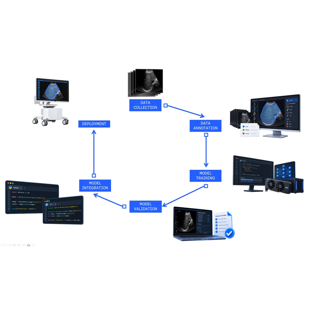
Medical imaging AI
We combine deep knowledge of medical imaging data with experience across segmentation, classification, detection, tracking, registration, and quantitative analysis. This allows us to to select and develop approaches suited to your specific clinical and technical challenge and to automate many steps of your workflow.
Data curation and annotation
Over the years, we have collected and curated numerous medical imaging datasets across a wide range of applications. We support dataset design, annotation workflows, and annotation bootstrapping, including efficient labeling with ImFusion Labels or other tools. We can also customize and fine-tune our off-the-shelf models for your data, helping you achieve strong performance with less development effort.
Model integration and deployment
We understand how model architecture, training data, validation methods, and computing constraints affect performance. Our extensive experience with AI frameworks, inference engines, and deployment environments enable us to integrate models in the most efficient way, whether it runs in the cloud or on surgical devices.
Product and regulatory readiness
We understand the traceability, reproducibility, validation, and documentation requirements for AI in regulated medical products. With that, we anticipate certification needs early and avoid costly changes later in development.
Multi-modal image registration
Align images across modalities, time points, and viewpoints
We are experts in state-of-the-art multi-modal registration that combines intensity-based, feature-based, and learning-based methods to align images acquired with different technologies, at different times, or from different viewpoints. The main challenges include differences in contrast, resolution, field of view, acquisition geometry, patient position, and tissue deformation.
We combine deep registration expertise with GPU-accelerated algorithms, automatic and customizable initialization, rigid global registration, and deformable models. Our platform supports 2D, 3D, surface, landmark, and point-cloud data, allowing us to build complete registration workflows across CT, MRI, ultrasound, X-ray, and endoscopic imaging.
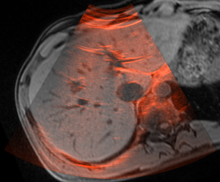
Multi-modal alignment
We developed algorithms that can robustly deal with differences in image contrast, resolution, field of view, and acquisition geometry. This allows us to align data any modality, including CT, MRI, Ultrasound, X-ray, PET, and endoscopic imaging.
Rigid and deformable registration
We model registration with the combination of rigid, affine, non-linear and dense deformation that better reflects how anatomy and imaging conditions change between images. This enables us to account for patient positioning, tissue deformation, anatomical movement and any change cause by the imaging setup.
2D, 3D, and surface registration
We combine image volumes, projection images, surfaces, landmarks, and point clouds across 2D/2D, 3D/3D, and 2D/3D workflows. This supports applications ranging from longitudinal analysis to intraoperative guidance.
Performance and workflow integration
We bring experience in GPU acceleration, automatic initialization, and learning-based registration. This helps you achieve reliable alignment within the performance and usability requirements of your clinical application.
Computational anatomy
Identify, model, and understand anatomy in medical images
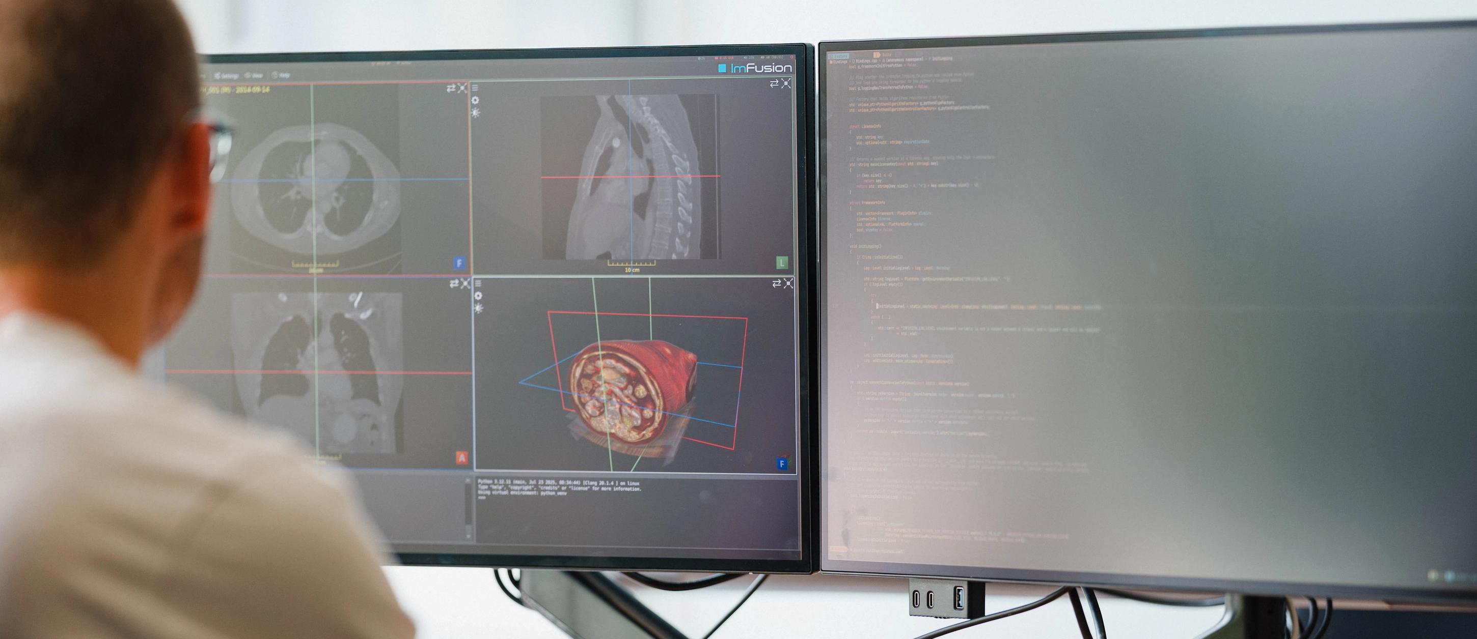
We develop methods that accurately detect and segment anatomical structures and clinically relevant pathologies in medical images. These are represented using data structures that draw from our experience in clinical applications in structured data combining segmentation labels, surfaces, landmarks, as well as metadata. Our primary focus is in working with X-Ray, CT, MRI, and Ultrasound images. These images come from abdominal, cardiac, orthopaedic, spinal, and cranial applications. The results from our libraries can directly be used in measurements, surgical planning, image registration, visualization, and navigation.
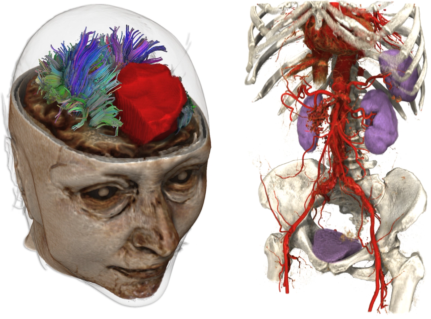
Anatomy-specific image analysis
We understand the anatomical variation and imaging characteristics that make automated analysis challenging and are aware of the typical challenges that might otherwise stop a naive approach in its tracks. This allows us to quickly develop robust approaches relevant to your application.
Structured anatomical representation
We combine label maps, surface meshes, landmarks, and relationships between multiple structures in coherent anatomical models. This gives you consistent, reusable data for downstream tasks such as measurements, planning, visualization, and navigation. Our architecture is built in a modular way that allows you to create complex pipelines by chaining together different independent steps.
Modality- and application-specific solutions
We have experience adapting anatomical analysis to different modalities and clinical fields, including CT and MRI applications for abdominal, cardiac, orthopedic, spinal, and cranial anatomy. In many cases, we have anatomy-specific libraries that can be integrated into a product. This enables us to shape the analysis around your data, workflow, and product requirements.
Real-time streaming
Acquire, process, and display imaging data as it is generated
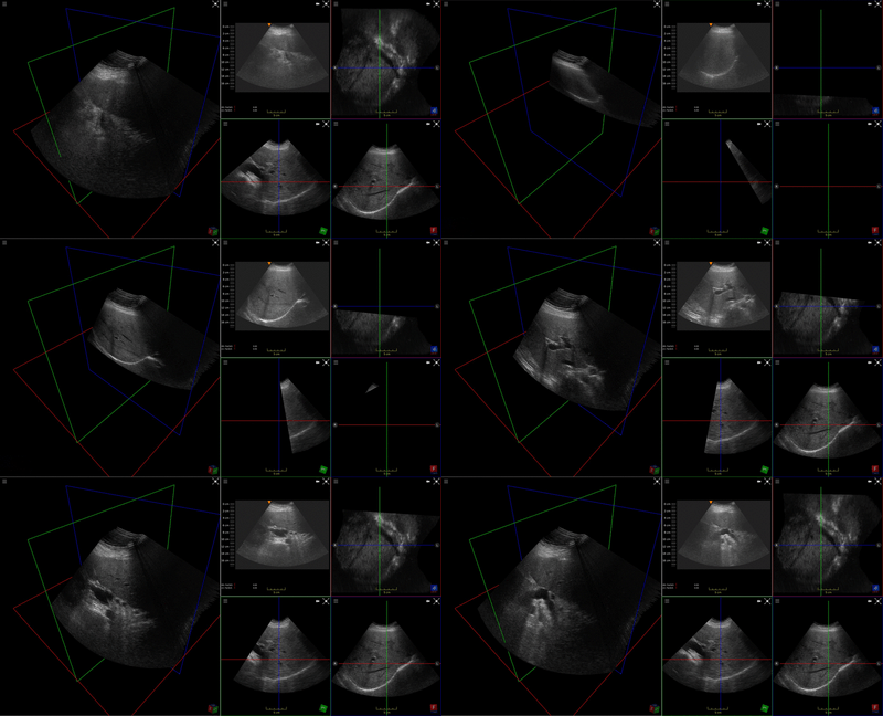
We build software pipelines that continuously receive imaging, video, tracking, and sensor data from medical devices. Our experience covers hardware integration, data synchronization, real-time processing, visualization, recording, playback, and network interoperability. This supports applications such as ultrasound guidance, endoscopic video analysis, surgical navigation, and robotic procedures.
Synchronization and data handling
We manage streams with different formats, frame rates, timestamps, and latencies. This keeps images, tracking information, and sensor measurements correctly aligned throughout the workflow.
Processing and visualization
We apply image processing, AI models, reconstruction, and registration directly to incoming data. GPU acceleration supports responsive visualization and analysis during time-critical procedures.
Recording, playback, and interoperability
We record one or several streams together, including mixed image and tracking sources, and replay them with original timing so the same pipelines run live or offline. The same stack connects local cameras and medical devices and exchanges streams with external systems over OpenIGTLink, network protocols, and ROS.
Robotics assisted workflows
Advanced robotic imaging and navigation
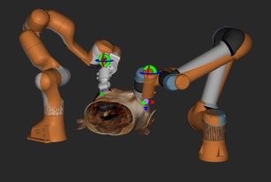
Medical robots need to operate as part of a wider clinical setup rather than as isolated devices. We combine medical imaging, computer vision, tracking, spatial registration, and robot control to help customers develop robot-assisted workflows that operate within defined clinical, technical, and safety constraints. This supports applications such as robotic ultrasound, image-guided interventions, automated instrument handling, and surgical navigation.
Robot and sensor integration
We connect robotic systems with cameras, tracking devices, imaging equipment, and force or tactile sensors. This provides the robot with the spatial information required to interact with the patient, instruments, and its environment.
Calibration and spatial registration
We determine the geometric relationships between robots, sensors, images, tools, and anatomical structures. This allows planned actions in medical images to be transferred accurately to the physical setup.
Robot control and motion planning
We work with robot kinematics, trajectory planning, collision checking, and constrained movement. This helps you develop safe and predictable robot behaviour for the available workspace and clinical task.
Workflow orchestration
We combine imaging, perception, robot control, and information about the current procedural step in one software architecture. This allows robotic actions to respond to the clinical workflow rather than operating as isolated movements.
Surgical guidance & navigation
Connect surgical plans with patient information, instruments, and live imaging
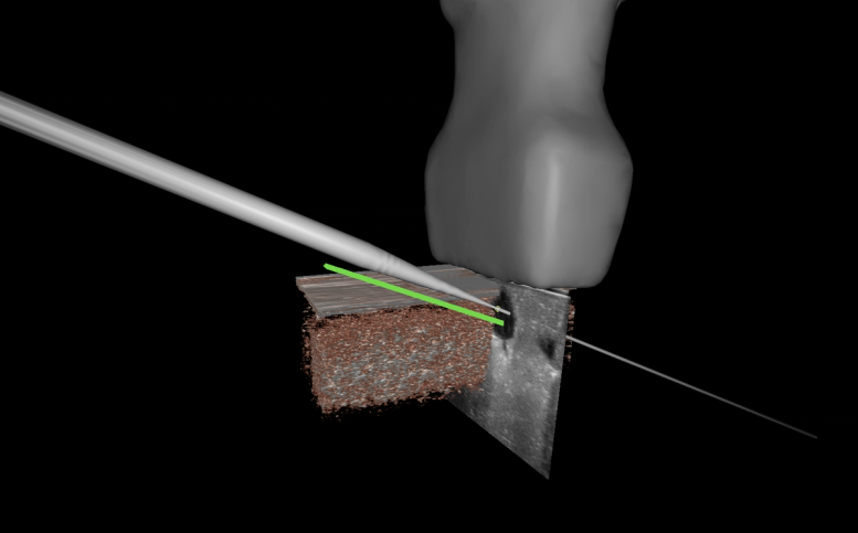
Surgical navigation shows the position of tracked instruments in relation to the patient’s anatomy and the planned procedure. We understand the complete spatial chain required to make this work, from patient registration and device calibration to real-time visualization and guidance. Our experience covers different tracking technologies, imaging modalities, and interventional workflows.
Tracking system integration
We integrate optical, electromagnetic, robotic devices, and other sensors providing real-time positional data with medical instruments, imaging devices, and software applications. This allows you to combine live position data with the anatomical and procedural information needed during an intervention.
Calibration and patient registration
We determine the spatial relationships between trackers, instruments, imaging devices, and the patient. Landmark-, surface-, and image-based registration methods connect preoperative plans with the physical anatomy in the operating room.
Navigation and guidance visualization
We develop 2D and 3D visualizations that show instruments, anatomy, targets, trajectories, and safety-relevant structures together. Guidance views such as tunnel, bullseye, and tool-oriented displays can be adapted to the procedure and user needs.
Integration of planning, imaging, and tracking
We combine preoperative plans, intraoperative images, and live tracking data in a shared 3D scene. This gives you a consistent spatial basis for navigation throughout the procedure.
Discuss your imaging goals, and constraints, then outline a clear path from concept to delivery.
Discovery Call
In a discovery call, we learn about your clinical or technical challenge and discuss how ImFusion’s expertise and technology could help make your idea a reality. Together, we identify promising first steps and determine where we can add the most value.
- Identify technical risks and regulatory considerations early
- Align engineering, clinical, and business stakeholders quickly
- Get a realistic timeline and resource estimate
- Leave with actionable next steps, not vague promises