Cranial Module
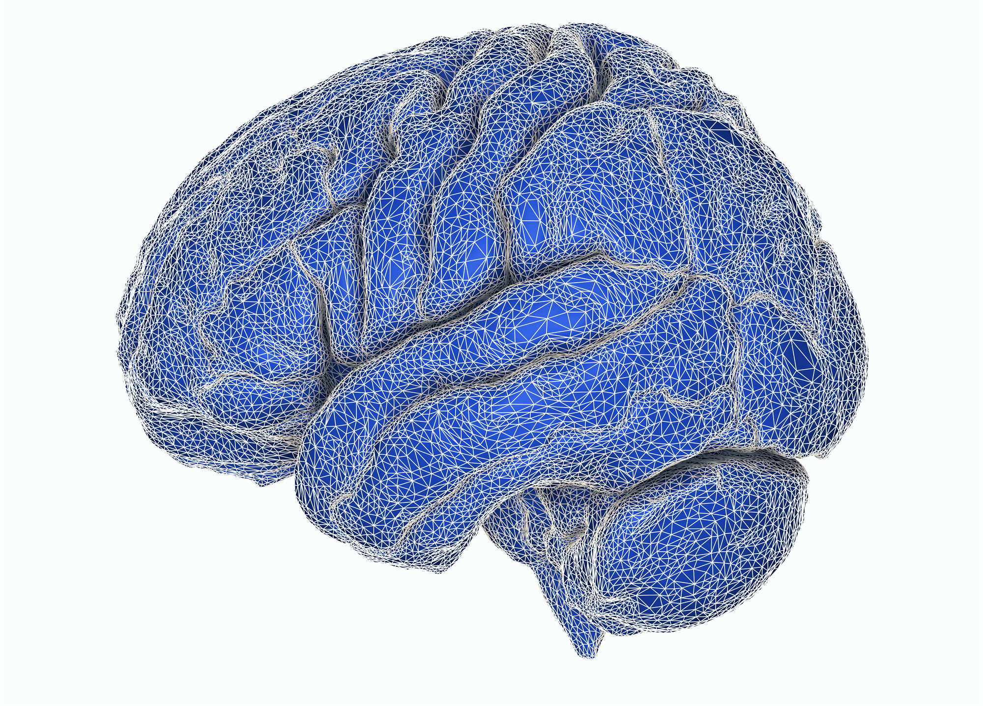
Gain full anatomical insights from your structural neuroimaging
Successful image-guided neurosurgery depends on turning raw CT and MRI into a precise, navigable 3D model — fast enough for the OR. The ImFusion Cranial module is a modular SDK component that puts segmentation, landmark detection, and multi-modal fusion in one framework, so each step feeds the next without stitching tools together.
A navigable 3D model from raw CT and MRI
We help you build tailored cranial pipelines on a shared anatomy model: bone from CT, brain and vessels from MRI, and a reproducible stereotactic frame. That model is ready for planning, fusion, and intra-operative guidance.
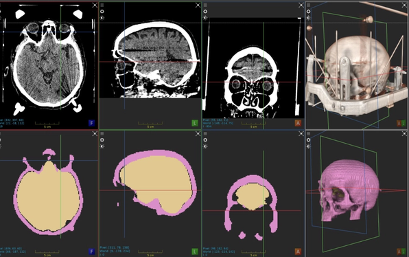
Cranial bone detection on CT and brain masking on MRI
We detect cranial bone on CT and mask the brain on MRI so later algorithms run on the anatomy that matters. On MRI, skull stripping produces a brain mask as a first step for parcellation and vessel work. On CT, we segment skull and craniofacial bone as a mask and mesh, and remove tables and fixtures, so the skull can constrain fusion and mark safe corridors.
- Brain masking on T1, T2, FLAIR, or T1CE, with optional mesh and crop
- CT skull and craniofacial bone as label map and mesh
- A first step for parcellation, fusion, and surgical corridors
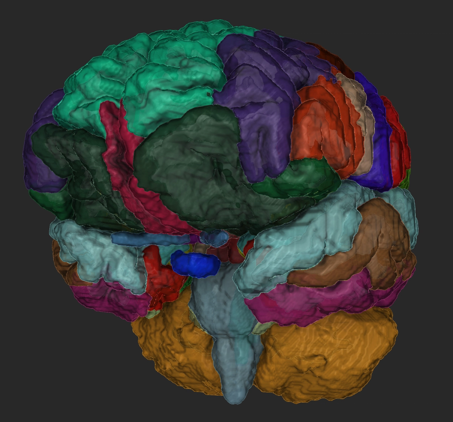
Brain parcellation on MRI
We parcellate the whole brain on T1-weighted MRI into a coherent map of cortical and subcortical regions. Optional meshes are grouped as named structures in the shared anatomy model, ready for planning, fusion, and visualization — without standing up a separate runtime.
- Whole-brain parcellation on T1-weighted MRI
- Cortical and subcortical regions as a coherent label map
- Optional named meshes for planning and visualization
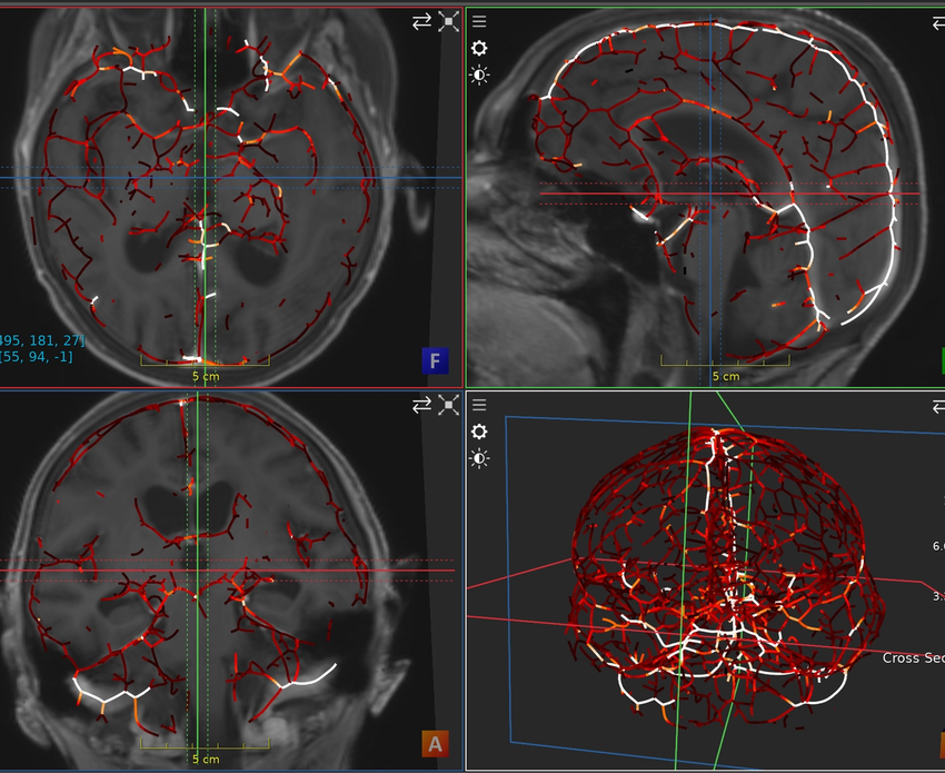
Vasculature segmentation
We segment cerebral vessels on contrast-enhanced MRI, typically T1CE, and can restrict detections to the brain. The result is a vessel map you can fuse with bone and parcellation, and from which we can extract centerlines and diameters when you need them. This algorithm is not thoroughly validated yet — it is under active development, and we can train it on your data together.
- Cerebral vessel segmentation on contrast-enhanced MRI
- Optional restriction to the brain mask
- Under active development; we can train on your data
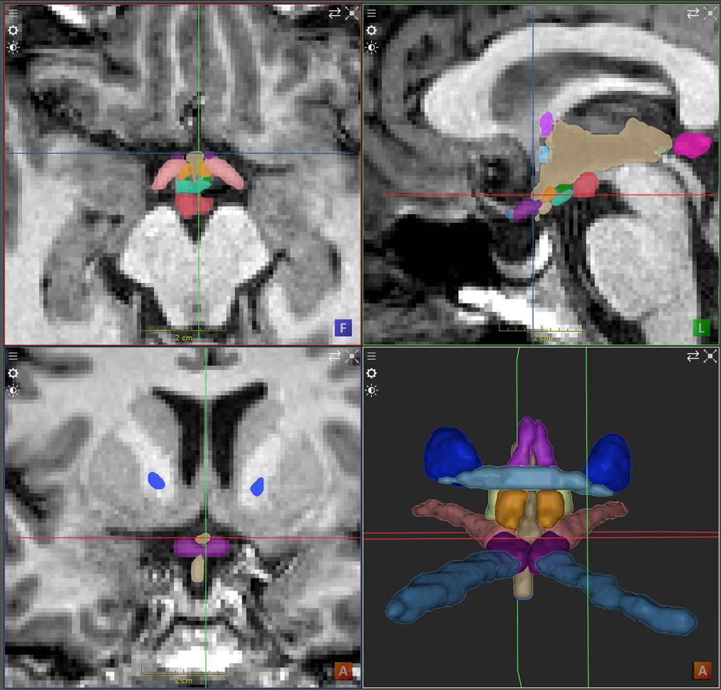
Landmarks and a shared anatomy model
Anterior and posterior commissure detection determine the standard stereotactic frame of reference on MR sequences T1, T1CE, T2, or FLAIR. Sequence classification identifies those contrasts automatically such that multi-modal pipelines can configure themselves. Each result form a single anatomy model, so the next algorithm, registration, or view does not start from raw files.
- AC and PC landmarks for stereotactic alignment
- Automatic MRI sequence classification (T1, T1CE, T2, FLAIR)
- One named anatomy model for maps, meshes, and fiducials
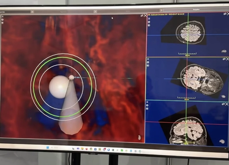
From fusion to neuronavigation
Our registration framework works on those anatomical outputs: rigid CT–MR fusion, atlas-to-patient alignment, and longitudinal MR–MR tracking. The CT skull can initialize the alignment; image-based refinement tightens soft tissue. High-performance rendering blends bone, MRI, and labels in real time — the same stack we use in neuronavigation workflows such as NousNav.
- CT–MR fusion for stereotactic planning and DBS
- Atlas-to-patient and follow-up MR–MR alignment
- Real-time fused visualization for neuronavigation
Get a technical walkthrough of the platform to see our key workflows in action and assess fit.
Product Demo
In a product demo, we show you how the ImFusion Suite, SDK, and ImFusion Labels support medical imaging and data annotation workflows. We focus on the capabilities relevant to your application and discuss directly how our technology could fit into your product or support your use case.
- See integration points mapped to your specific tech stack
- Identify bottlenecks before committing development resources
- Validate data format compatibility and transformation requirements
- Get immediate answers from engineers who built the system