Guiding cardiac interventions with real-time heart models
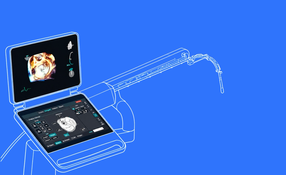
How ImFusion is enabling intelligent ultrasound guidance for robotic probe navigation
Project overview
A medical device company is developing a robotic system for navigating transesophageal echocardiography probes during procedures such as valve repair and left atrial appendage closure. ImFusion contributes real-time anatomical analysis and spatial guidance that help the system understand what is visible in the ultrasound image and how the probe is positioned relative to the heart.
- Project type: Industry collaboration
- Clinical field: Interventional cardiology
- Expertise applied: Ultrasound / machine learning / anatomical modelling / real-time processing / registration / visualization
- Project state: Product development
Keeping the right anatomy visible throughout a beating-heart procedure is a demanding task
The challenge
Transesophageal echocardiography (TEE) provides only a limited view of the heart at any given moment. The system must identify the visible anatomy despite image artifacts and constant cardiac motion, determine the current imaging plane, and provide information that can support safe robotic probe movements.
- Interpret volumetric TEE images in real time
- Recognise anatomy within a limited field of view
- Account for imaging artifacts and the beating heart
- Determine the probe’s position relative to the heart
Our approach
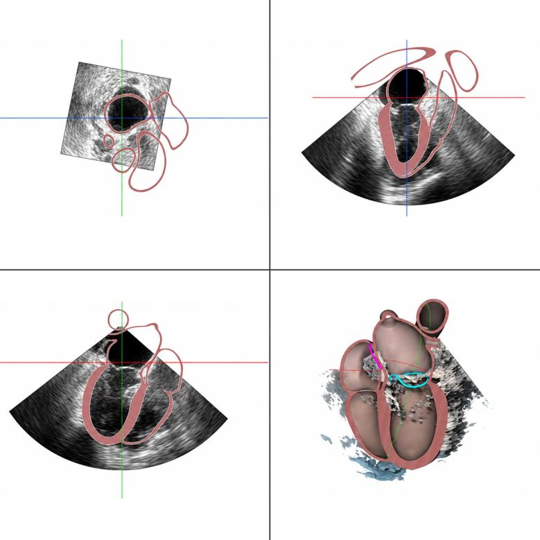
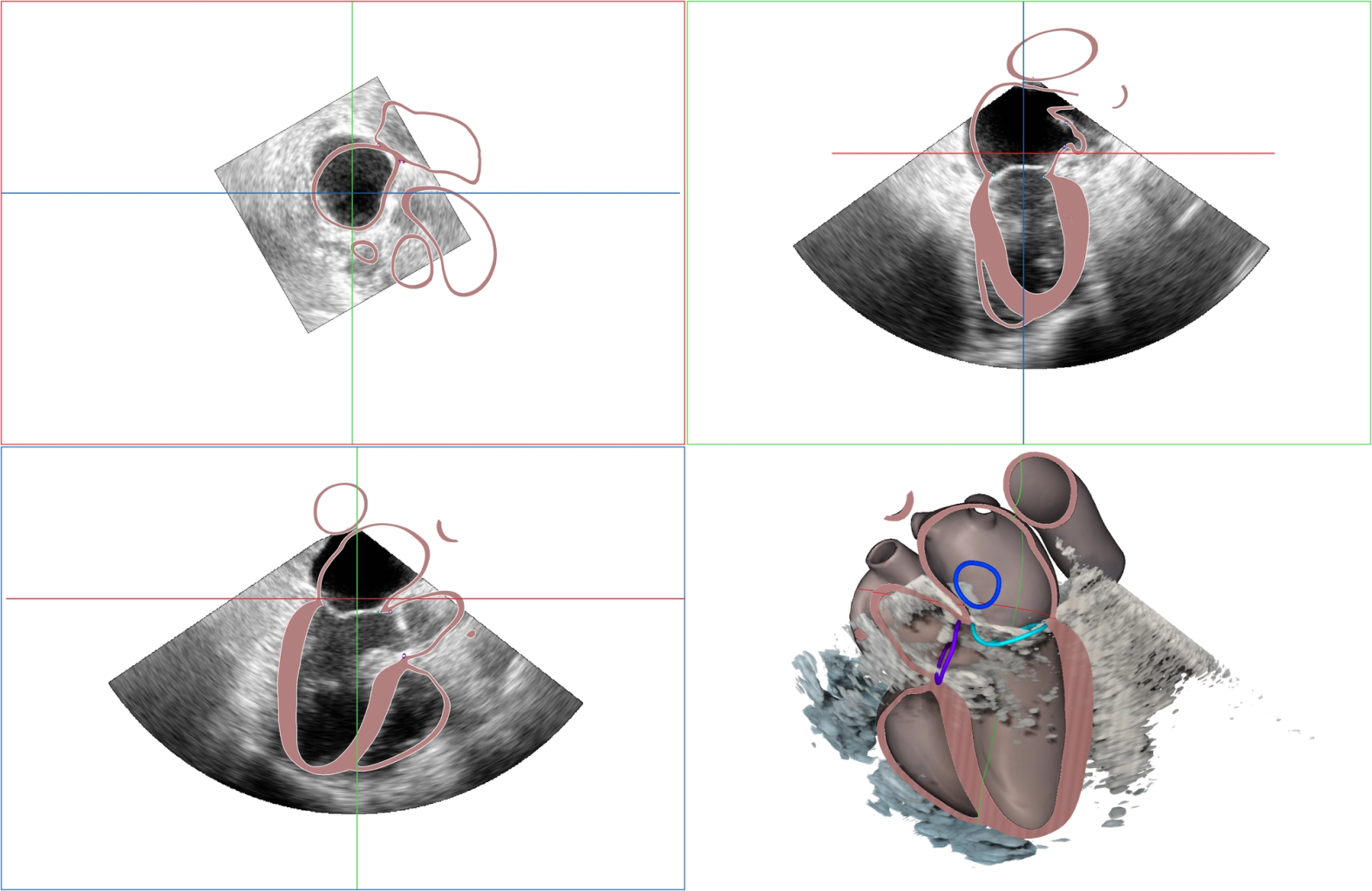
Instead of analysing only the anatomy currently visible in the ultrasound image, ImFusion used advanced AI to create a detailed, patient-specific 3D heart model that extends beyond the field of view. From the heart model, precise landmark locations can be derived to localize anatomical structures and view planes. Also, the model is used to track the probe relative to the heart at any given moment.
- Fit a volumetric heart model to live TEE data
- Represent anatomy beyond the visible image region
- Estimate the relationship between probe and heart
- Provide spatial information for robotic probe guidance
The impact
Reliable anatomical understanding is a fundamental requirement for robotic ultrasound guidance. Maintaining consistent spatial context throughout the procedure could make demanding imaging tasks easier and reduce the need for a second clinician dedicated solely to probe handling.
- Support consistent visualization during an intervention
- Provide spatial information for robotic navigation
- Reduce the technical burden on the clinical team
Our process
Build the training data
We used ImFusion Labels to create detailed training data from cardiac ultrasound volumes. The annotations included anatomical segmentations and geometric information required for model development.
Develop the anatomical model
We developed graph-based machine-learning methods that fit a strong anatomical model of the heart to volumetric TEE images. The model provides a structured representation of the patient’s anatomy rather than an isolated segmentation.
Iterate and refine
Several iterations of dataset preparation, preprocessing, model architecture, and downstream processing were required. Interactive prototypes allowed the team to assess results early and refine the approach throughout development.
Connect analysis and guidance
The anatomical model was integrated with real-time image processing and 2D and 3D visualization. Its outputs provide landmarks, imaging planes, and spatial information for integration into a robotic probe navigation system.
Four-dimensional ultrasound provides rich information about cardiac anatomy and motion, but the challenge is to show clinicians only what they need at each stage of the procedure.
ImFusion technology
ImFusion contributed to the imaging and analysis pipeline, from training-data preparation and interactive prototyping to real-time processing and visualization. ImFusion Labels supported data annotation, while the ImFusion Suite and SDK provided the basis for rapid experimentation and integration into the developing system.
ImFusion expertise
The project brings together our experience in cardiac ultrasound, anatomical modelling, machine learning, data curation, real-time image processing, visualization, and medical-software development. This combination allows us to connect individual technical components into an imaging workflow designed around an interventional use case.
We offer two types of introductory calls so we can make sure we’re speaking to the right audience.
Product Demo
In a product demo, we show you how the ImFusion Suite, SDK, and ImFusion Labels support medical imaging and data annotation workflows. We focus on the capabilities relevant to your application and discuss directly how our technology could fit into your product or support your use case.
- See integration points mapped to your specific tech stack
- Identify bottlenecks before committing development resources
- Validate data format compatibility and transformation requirements
- Get immediate answers from engineers who built the system
Discovery Call
In a discovery call, we learn about your clinical or technical challenge and discuss how ImFusion’s expertise and technology could help make your idea a reality. Together, we identify promising first steps and determine where we can add the most value.
- Identify technical risks and regulatory considerations early
- Align engineering, clinical, and business stakeholders quickly
- Get a realistic timeline and resource estimate
- Leave with actionable next steps, not vague promises